| Company name | Hamamatsu Photonics | Roche Diagnostics Deutschland GmbH | Sysmex Deutschland GmbH 3DHISTECH LTD | |||
|---|---|---|---|---|---|---|
 | 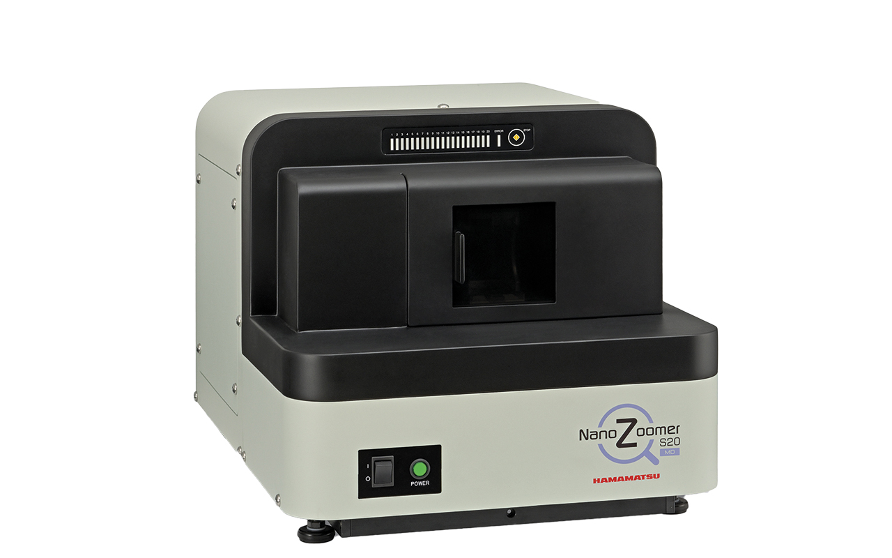 | 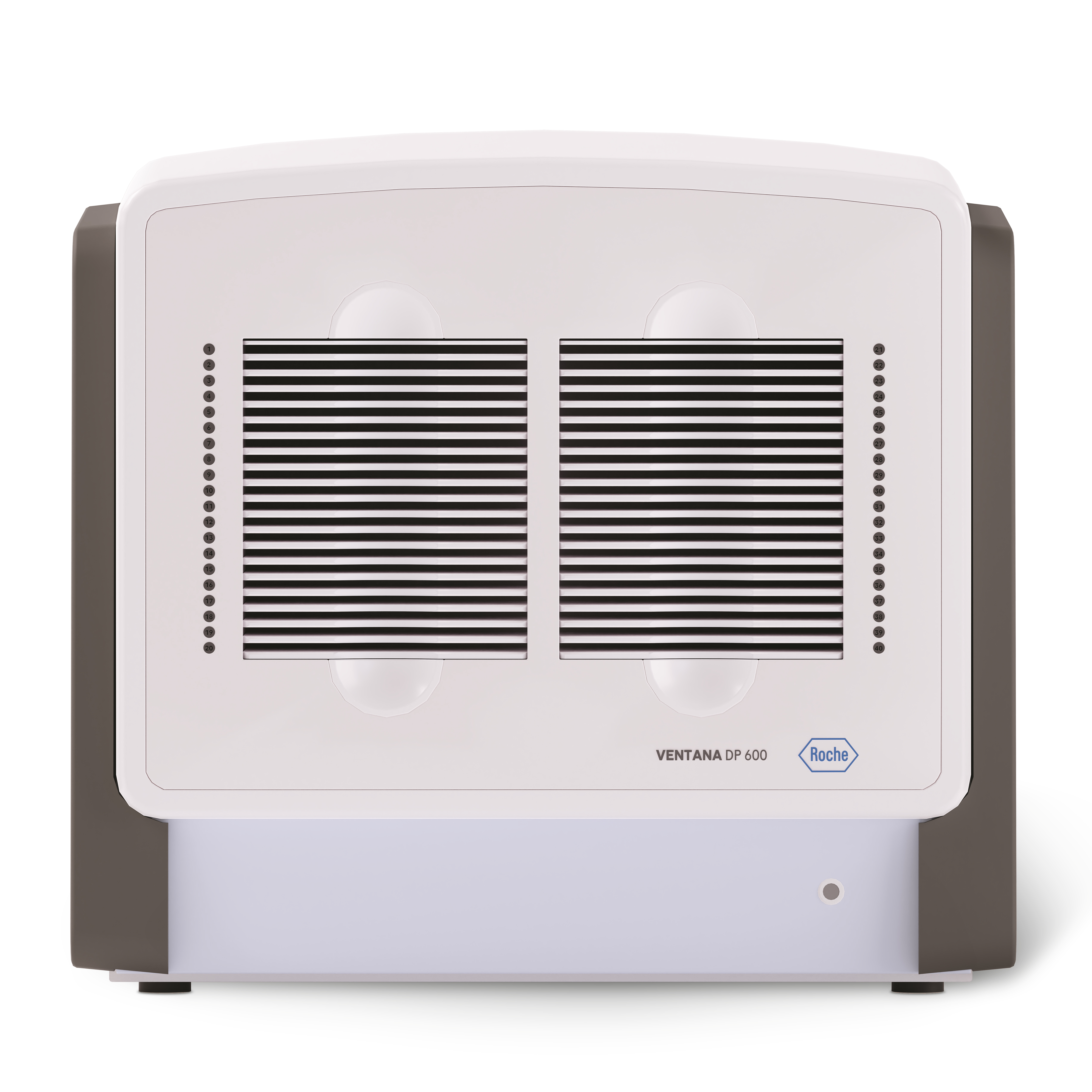 | 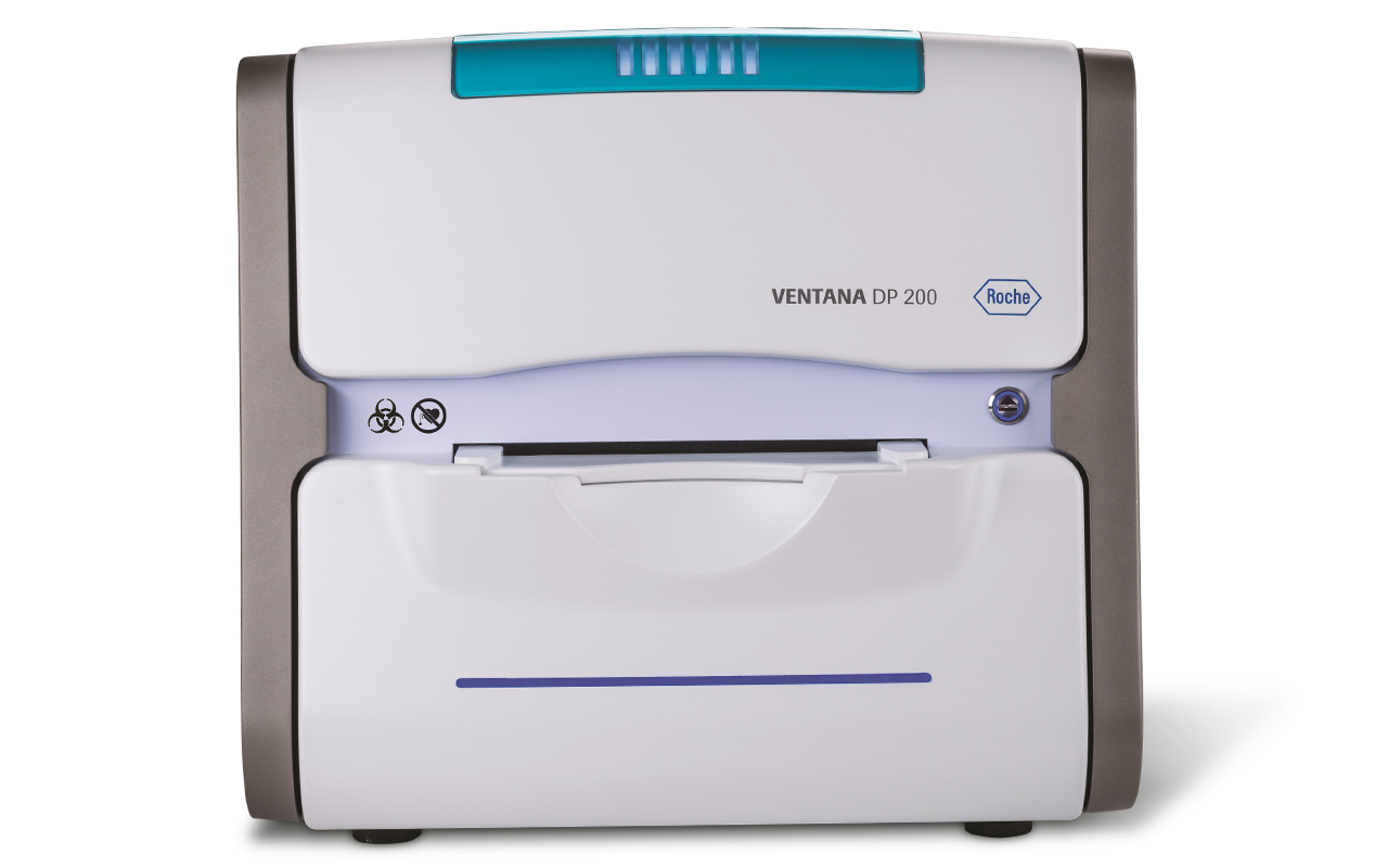 | 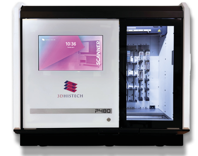 | ||
| Model name | NanoZoomer S540MD | NanoZoomer S20MD | Ventana DP600 | Ventana DP200 | Pannoramic 480 2nd Gen. | |
| System data | Type of construction | Benchtop or placed on cart | Benchtop | Integrated, compact benchtop device | Integrated, compact benchtop device | Integrated, benchtop |
| Width x height x depth (cm) | 75 x 64 x 69 | 48 x 45 x 61 | 74 x 74 x 67 | 50 x 68 x 46 | 119 x 100 x 90 | |
| Weight (kg) | 123.6 (188.6 with cart) | 52 | 75 + 11 (Touchscreen Monitor + PC) | 46,5 + 11 (Touchscreen Monitor + PC) | 240 | |
| Supported slide dimensions (width x length x thickness in mm) | 25–26 x 75–76 x 0.9–1.2 | 25–26 x 75–76 x 0.9–1.2 | 25–26 x 75–76.5 x 0.9–1.4 | Single: 25–26 x 75–76.5 x 0.9–1.4 Double: 51–52 x 75–76.5 x 0.9–1.4 | Single: 25–26 x 75–76 x 0.9–1.2 Double: 50–51 x 75–76 x 0.9–1.2 | |
| Slide handling | Automated, horizontal | Automated, horizontal | Horizontal, slide remains in the tray (exclusion of glass breakage) | Automated, horizontal | ||
| Lenses (magnification | numerical aperture) | 20x | 0.75 | 20x | 0.75 | 20x | 0.75 (Nikon ApoChromat, WD = 1.0 mm, additional LS for 40x) | 20x | 0.8; 40x | 0.95; 40x | 1.2 (water immersion) | ||
| Resulting magnification per lens (x-fold) | 20x; 40x | 20x; 40x | 20x; 40x | 20x; 40x | Up to 57x; 113 x; 113x (depending on camera type) | |
| Pixel resolution (µm/pixel) per lens | 20x: 0.46; 40x: 0.23 | 20x: 0.46; 40x: 0.23 | 20x: 0.46; 40x: 0.25 | 20x: 0.46; 40x: 0.25 | 0.18; 0.09 (depending on camera type) | |
| Digital image sensor (type | pixel MP) | CMOS | – | CMOS | – | CMOS Tri-Linear | 24 | CMOS Tri-Linear | 24 | CMOS | 12; 21; 25 (depending on configuration) | |
| Image processing | Automatic white balance, auto focus | Automatic white balance, auto focus | Automatic white balance, ICC color profiles, dynamic autofocus, AI-supported tissue recognition | Automatic white balance, auto focus, color calibrated | ||
| Supported microscopy methods | Brightfield microscopy | Brightfield microscopy | Brightfield microscopy | Brightfield microscopy | Brightfield microscopy, polarization (optional) | |
| Light source types | LED | LED | LED | LED | Pulsed xenon lightsource | |
| Performance data | Slide loader (type | loading capacity) | 540 slides | 30 slide cassettes; 360 slides | 20 slide cassettes | 20 slides | 1 cassette | Slide tray | 40 trays with 6 slides each (240 in total) | Slide tray | 1 tray with 6 slides | Racks | up to 480 slides |
| Supported stainer racks (type) | Sakura 4768 20-slide cassette; Leica Universal 30-slide cassette | Sakura 4768 20-slide cassette; Hamamatsu Slide cassette | – | – | Sakura, Leica | |
| Image formats (incl. conversion) | NanoZoomer Digital Pathology Image (NDPI), DICOM | NanoZoomer Digital Pathology Image (NDPI), DICOM | BIF, TIF, DICOM | BIF, TIF, DICOM | Native: MRXS, DICOM, export to svs or tiff | |
| Z-stacks (number | distance) | 1–99 layers | up to 100 μm | 1–99 layers | up to 100 μm | 15 | 0.5–1.5 μm | 30 | 0.2–10 µm | ||
| Scanning speed for an area of 15 x 15 mm (enlargement I sec.) | 20x; 40x | approx. 30 sec (area of 15 mm × 15 mm square with 5 focus points) | 20x | 36 sec; 40x | 73 sec | 40x | approx. 30 sec | |||
| Throughput: slides/h | ≥ 82 | 80 | 20x: approx. 70 (time to view); 40x: approx. 40 (time to view) | 80 | ||
| Integrated QC and Indication | Yes, manual QC or integrated focus score, with possibility of automatic rescan at specified score levels | Yes, manual or automatic advanced image quality check | Yes, manual image quality check | Internal QC for focus quality | ||
| Integration and Networking | Viewer | Stand-alone, included with NZAcquireMD software, or optional web-based server managed software for scanning and viewing | Slide Viewer (stand-alone), navify Digital Pathology IMS (web-based) | Stand-alone and web-based | ||
| AI-Support | Yes, intelligence mode tissue detection to detect areas of tissue on the slide macro image | Yes, with navify Digital Pathology IMS | Yes, AI supported tissue detection | |||
| Works with Cloud and/or onPrem | Cloud and on-prem storage | Cloud and on-prem storage | Slide Viewer (on-prem), navify Digital Pathology IMS (cloud) | On premise | ||
| Self-calibration | Automatic self-correction | Yes | Yes | Yes | Yes | |
| STAT mode, prioritised slides | Yes, prioritise and organise the scanning order of racks or slides. | Yes, although it is not required with a low capacity scanner you can define the order of scanning. | Yes, trays can be prioritized | – | Yes | |
| Barcode scanner (1D, 2D) | Barcode driven workflow with 1D and 2D barcode support | 1D, 2D: DATAMATRIX, PDF417, QR code | 1D, 2D: DATAMATRIX, PDF417, QR code | 1D, 2D | ||
| Image data management | Hamamatsu image formats are compatible with all major providers of image management software. We can also provide our NZConnect software for management of images. | IVD-compliant IMS for diagnostics (also across locations): navify Digital Pathology | RX: SlideCenter; DX: CaseManager | |||
| Interface protocols | Supports HL7 interfacing and automated DICOM image distribution to IMS, PACS, and VNA for efficient data exchange | HL7, DICOM | HL7, DICOM | HL7 | ||
| Connection to LIS/PIS | HL7 interfacing for LIS and PIS integration, automated LIMS data association via barcode recogni-tion, bidirectional communication with LIS/PIS, NZConnect API for custom LIS/PIS integration. | Yes | Yes | Yes | ||
| Certifications | Compliant with IVDR, IvDO and UK MDR2002 | CE-IVD, FDA | CE-IVD, FDA | DX: IVDR | ||
| DICOM supported | Yes | Yes | Yes | Yes | Yes | |
| Embeds color profile | Embedded CCM | Embedded CCM | ICC color profiles | ICC color profiles | Yes | |
| Software development kit (SDK) available | Yes | Yes | On request | On request | Yes, 3DHistech SimpleSlideInterface | |
| Unique selling points, special features | The NanoZoomer S540MD is a large capacity, high-throughput, automated scanner that seamlessly integrates to lab environments. With Sakura cassettes support, touchscreen interface it is intuitive requiring minimal user input. | The NanoZoomer S20MD is a high-speed, compact, and adaptable scanner that adds flexibility and scalability to your digital pathology workflow. It supports Sakura cassettes and automatic scanning for minimal user input. | Medium to high throughput in histopathological routine diagnostics, CE-IVD system, can be combined with the IMS navify Digital Pathology and thus access to a variety of algorithms is possible | Scanning of double-width slides is possible, ideal for frozen section application, CE-IVD system, suitable for cytology and histopathology, combinable with the IMS navify Digital Pathology and thus access to a variety of algorithms is possible | With all-in-one Server PC with up to 40 GB storage, only high-troughput scanner with 40x objective, water immersion or polarization mode, RX package includes IMS (SlideCenter) and quantification software, DX package in-cludes IMS (case manager), viewer and diagno-stic applications AI and monitoring software | |
| Contact | Hamamatsu Photonics Deutschland GmbH Erk Klopp +49 8152 375 212 erk.klopp[at]hamamatsu[dot]eu www.hamamatsu.de | Roche Diagnostics Deutschland GmbH Julia Schiffer +49 173 586 1010 julia.schiffer.js1[at]roche[dot]com www.roche.de | Sysmex Deutschland GmbH Olaf Scheel +49 174 378 7092 scheel.olaf[at]sysmex[dot]de www.sysmex.de | |||
Pathology