Viewer Software | VMscope GmbH kai.saeger@vmscope.de | Roche Diagnostics Deutschland GmbH mannheim.digitalprodukte@roche.com | Sysmex Deutschland GmbH Bornbarch 1, 22848 Norderstedt, Germany, Telefon: +49 40 534 102 0 info@sysmex.de www.sysmex.de/digitalepathologie | |||
|---|---|---|---|---|---|---|
|
|
| 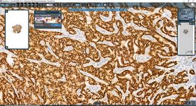 | 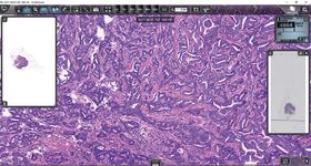 |  | |
URL | https://www.sysmex.de/produkte/details/clinical-viewer.html | https://www.sysmex.de/produkte/details/slideviewer.html | https://www.sysmex.de/produkte/details/instantviewer.html | |||
Product name | VM Slide Explorer | navify® Digital Pathology | Clinical Viewer | SlideViewer | InstantViewer | |
Software details | Description/Slogan | Digital microscope as local application | navify® Digital Pathology enhances the efficiency of pathology laboratory workflow with connectivity and automation. The cloud-based pathology workflow software seamlessly connects technicians and pathologists for communication and collaboration. navify® Digital Pathology is the Software as a Service version of Roche uPath enterprise software (est. 2019). | Clinical Viewer is a digital microscopy application designed for supporting the histopathological diagnostic workflow and the microscope examination process. | SlideViewer is a digital microscopy application designed for supporting the microscope examination process in bioscience | InstantViewer is the default application for opening slides in SlideCenter. It is a multi-platform slide viewer application supporting the following platforms: Windows, Mac OSX, iOS, LINUX and android |
Established (continuous improvements and updates) | 2022 | 2022 | since 2020 | since 2013, current release: SlideViewer 2.6 | ||
Pricing Model | Client licenses | Pay per user, subscription | Integral part of Pannoramic Pathology Management System (PPMS) | free of charge | free of charge | |
Primary Use Case | Research use | Clinical, research, educational | Diagnostic | Research | Research, front end viewer of slide management system SlideCenter | |
Certificates | DIN EN ISO 9001:2016 and 13485:2021 certificate, no IVD certification yet | CE-IVD | CE-IVD (IVDR) | |||
Architecture | Windows application | Cloud based, server based, web application | Windows application | Windows application | Webbrowser based; also available as iPad Viewer app | |
Compatibility Scanner-/Fileformats | VMscope .vsf, 3DHistech Pannoramic .mrxs, Hamamatsu .ndpi, Aperio ScanScope .svs, Olympus WebImage, Leica .scn, Zoomify, JPEG2000 .jp2, BigTiff .tif, Precipoint .vmic, Philips .iSyntax, Zeiss .czi, Argos .avs | bif, tiff, svs, scn, ndpi, dicom | mrxs, svs, dicom, ndpi, iSyntax, tiff | mrxs, svs, dicom, ndpi, iSyntax, tiff | mrxs | |
Compatibility LIS (HL7 based LIS integration) | Nexus/Pathologie, DC-Systeme dc-pathos, BasysData PathoWin, Telemis, Kairos, Co-Fox | HL7 enables integration with all LIS | Yes | Yes | Yes | |
Application Programming Interface (API) | Yes | Yes | Yes | Yes | N/A | |
Telepathology function | Yes | Yes | Yes, via Pannoramic Pathology Management System (PPMS) | Yes, via SlideCenter | Yes, via SlideCenter | |
Handling and functions | Storage location (Cloud, local) | Local | Cloud and local | Server based storage (PPMS) | Both, local and cloud or server | Only slides on server/cloud |
Time required for saving process | Local streaming | Will save immediately to IMS after scanning | Instantly | Instantly | Instantly | |
Support sharing | Yes | Yes | Yes, via Pannoramic Pathology Management System (PPMS) | Yes, via SlideCenter | Yes, via SlideCenter | |
Supports screenshots | Yes | Yes | Yes | Yes | Yes | |
Includes AI tools | Ki67, ER, PR and HER2 with CE certificate, several further RUO modules | Yes (uPath image analysis algorithms and open environment for external algorithms for pathology decision support) | Integration of AI tools via python interface, image analysis tools included (IHC, tissue separation, MarkerCounter) | Integration of AI tools via python interface, Image analysis tools optional (QuantCenter) | N/A | |
Comments | Yes | Yes, comments can be added | ||||
Special features | Unique selling proposition, special features |
|
|
|
|
|